Chemokine guidance of central memory T cells is critical for antiviral recall responses in lymph nodes

Citation:

Sung JH, Zhang H, Moseman AE, Alvarez D, Iannacone M, Henrickson SE, de la Torre JC, Groom JR, Luster AD, von Andrian UH. Chemokine guidance of central memory T cells is critical for antiviral recall responses in lymph nodes. Cell. 2012;150 (6) :1249-63.

Date Published:

2012 Sep 14

Abstract:

A defining feature of vertebrate immunity is the acquisition of immunological memory, which confers enhanced protection against pathogens by mechanisms that are incompletely understood. Here, we compared responses by virus-specific naive T cells (T(N)) and central memory T cells (T(CM)) to viral antigen challenge in lymph nodes (LNs). In steady-state LNs, both T cell subsets localized in the deep T cell area and interacted similarly with antigen-presenting dendritic cells. However, upon entry of lymph-borne virus, only T(CM) relocalized rapidly and efficiently toward the outermost LN regions in the medullary, interfollicular, and subcapsular areas where viral infection was initially confined. This rapid peripheralization was coordinated by a cascade of cytokines and chemokines, particularly ligands for T(CM)-expressed CXCR3. Consequently, in vivo recall responses to viral infection by CXCR3-deficient T(CM) were markedly compromised, indicating that early antigen detection afforded by intranodal chemokine guidance of T(CM) is essential for efficient antiviral memory.
Cell
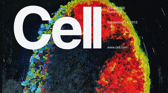
The ability to learn and remember previously encountered pathogens is a hallmark of the vertebrate immune system. CD8+ T cells, called central memory cells (TCM), mediate much more rapid and vigorous immune responses against the viruses that they recognize compared to their uneducated precursors, the naive T cells (TN). In this issue, Sung et al. (pp. 1249–1263) compare, at the single-cell level, the response of TN and TCM to a subcutaneous viral challenge. The invading virions are rapidly transported to the draining lymph nodes, where they infect macrophages that line the periphery of these bean-shaped organs. The image shows a cross-section of a lymph node in which virus-infected macrophages are identified by their yellow-green color. Initially, both TN and TCM reside in the deep T cell area (the dark region in the center of the organ). TCM, unlike TN, expresses CXCR3, a chemokine receptor that enables TCM to sense distant viral infections and to migrate peripherally between the B cell follicles (red) and into the medulla (the blue-green region on the left). This chemokine-dependent redistribution of TCM provides rapid access to viral antigen, which is critical for expedient clearance of the virus and, thus, represents a key molecular feature of immunological memory. Oil on canvas painting by Meghan Perdue.

Cover by Megan Perdue (oil on canvas)

Last updated on 03/31/2023