Photos
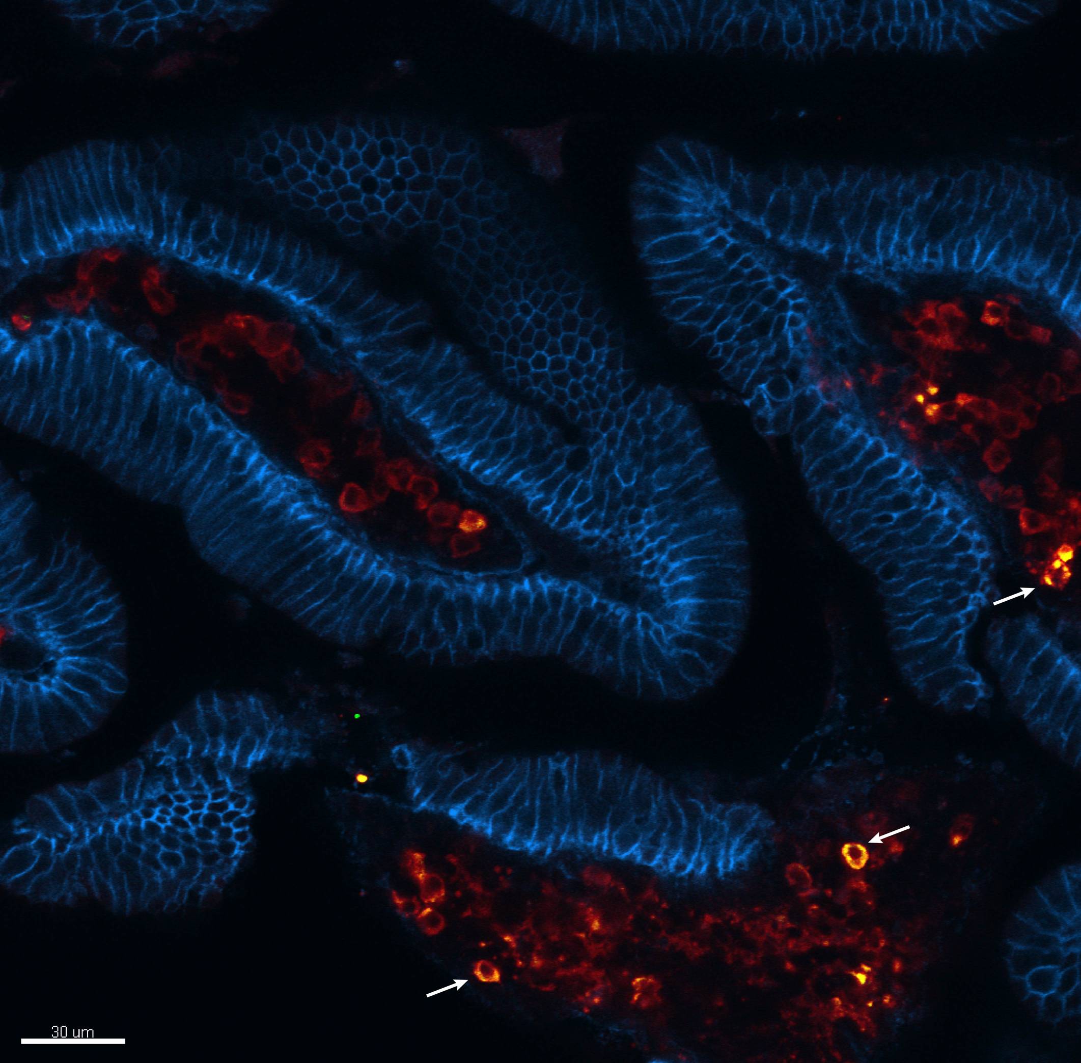
Small intestine villi (epithelium stain in blue) showing IgA producing cells (in red) with antigen specificity (yellow = green + red, indicated by white arrows), 5 months after parenteral immunization.
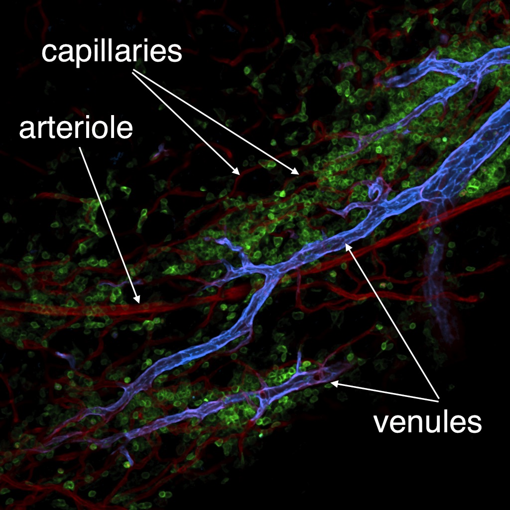
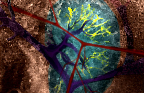
Whole-mount staining of mouse omentum. All microvessels, such as capillaries, venules and arterioles are labeled red with the pan-endothelial marker CD31, venules are labeled in blue with a monoclonal anti-DARC Ab developed in my lab. Hematopoietic cells are labeled in green with anti-CD45 Ab. Only venules expressing DARC are capable of efficient leukocyte recruitment from the blood.
Cover of Immunity (vol 45, issue 6, December 2016)
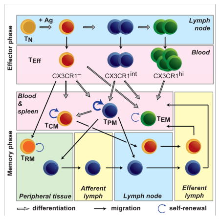
Chemokine Receptor CX3CR1 Defines Three Antigen-Experienced CD8 T Cell Subsets with Distinct Roles in Immune Surveillance and Homeostasis.
Summary Figure: Gerlach C, Moseman AE, Loughhead SM, Alvarez D, Zwijnenburg AJ, Waanders L, Garg R, de la Torre JC, von Andrian UH. The Chemokine Receptor CX3CR1 Defines Three Antigen-Experienced CD8 T Cell Subsets with Distinct Roles in Immune Surveillance and Homeostasis. Immunity. 2016;45 (6) :1270-1284.
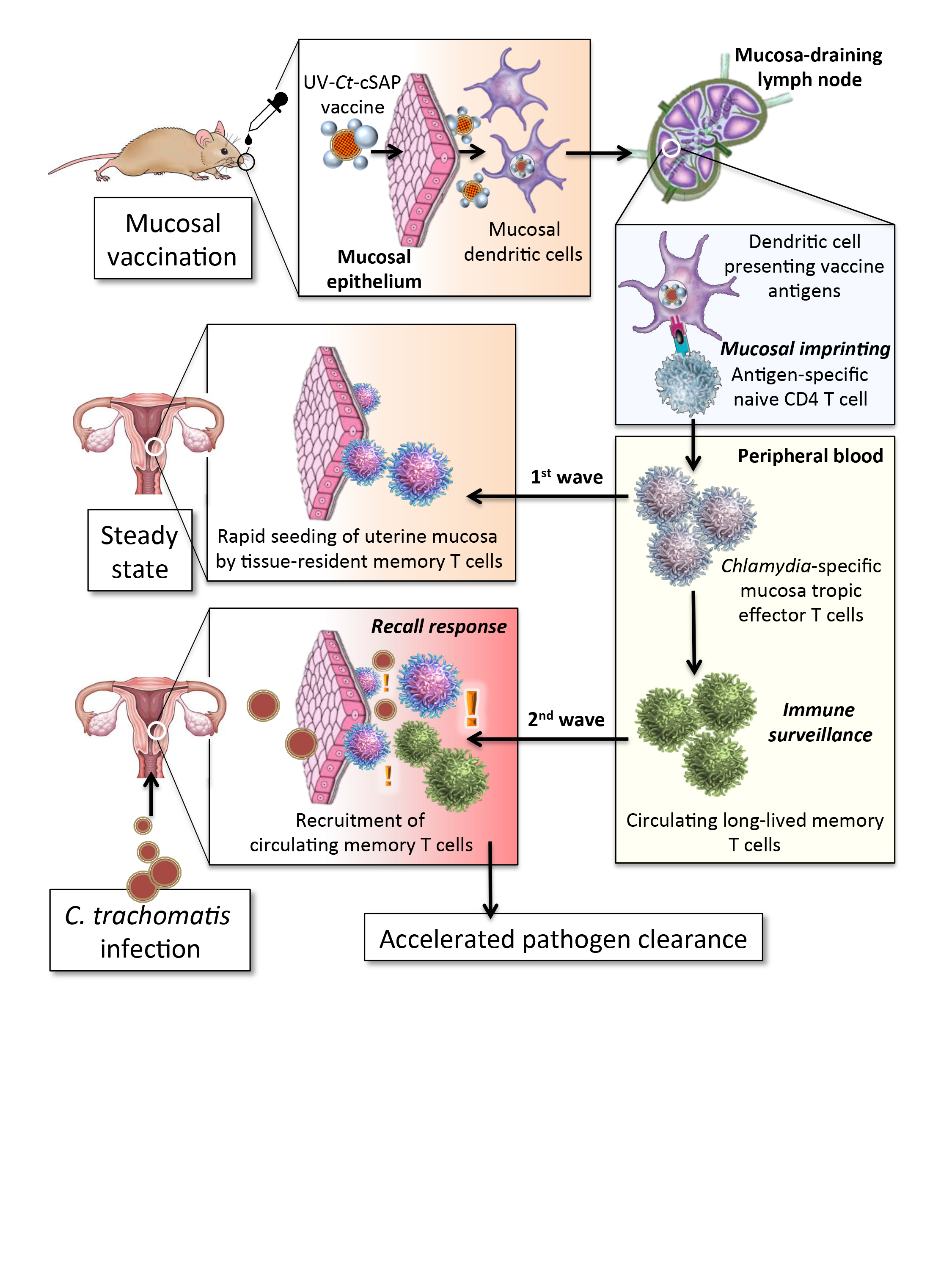
Protection against C. trachomatis infection after musocal UV-Ct-cSAP vaccination
Protection against C. trachomatis infection after musocal UV-Ct-cSAP vaccination Upon mucosal vaccination, dendritic cells carry UV-Ct-cSAP to lymph nodes and stimulate CD4 T cells. Effector T cells are imprinted to traffic to uterine mucosa (first wave) and establish tissue-resident memory cells (TRM cells). Vaccination also generates circulating memory T cells. Upon genital Ct infection, local reactivation of uterine TRM cells triggers the recruitment of the circulating memory subset (second wave). Optimal pathogen clearance requires both waves of memory cells.
Cover of Nature Nanotechnology (vol. 9, issue 8, August 2014)
Small interfering RNAs (siRNAs) can turn off any specific gene in the genome. As a result, these molecules have tremendous potential as both scientific tools and therapeutics. However, delivering siRNA to the right cells in vivo has remained challenging. Using combinatorial chemical synthesis techniques and high-throughput biological screening methods, Daniel Anderson and co-workers have designed a nanoparticle that delivers siRNA to endothelial cells — cells that line blood and lymphatic vessels — at very low doses. Using this nanoparticle, the researchers turned off five genes at once inside an animal, turned off genes for more than three weeks after one injection, and reduced inflammation, tumour growth and metastasis. The cover image shows blood vessels in mouse adipose tissue stained with two endothelial cell markers, CD31 (blue) and ICAM-2 (magenta). Cover Art by Aude Thiriot
Nociceptive sensory neurons
Summary Figure: Riol-Blanco L, Ordovas-Montanes J, Perro M, Naval E, Thiriot A, Alvarez D, Paust S, Wood JN, von Andrian UH. Nociceptive sensory neurons drive interleukin-23-mediated psoriasiform skin inflammation. Nature. 2014;510 (7503) :157-61.
Cover of Cell (vol. 150, issue 6, Sept 14, 2012)
The ability to learn and remember previously encountered pathogens is a hallmark of the vertebrate immune system. CD8+ T cells, called central memory cells (TCM), mediate much more rapid and vigorous immune responses against the viruses that they recognize compared to their uneducated precursors, the naive T cells (TN). In this issue, Sung et al. (pp. 1249–1263) compare, at the single-cell level, the response of TN and TCM to a subcutaneous viral challenge. The invading virions are rapidly transported to the draining lymph nodes, where they infect macrophages that line the periphery of these bean-shaped organs. The image shows a cross-section of a lymph node in which virus-infected macrophages are identified by their yellow-green color. Initially, both TN and TCM reside in the deep T cell area (the dark region in the center of the organ). TCM, unlike TN, expresses CXCR3, a chemokine receptor that enables TCM to sense distant viral infections and to migrate peripherally between the B cell follicles (red) and into the medulla (the blue-green region on the left). This chemokine-dependent redistribution of TCM provides rapid access to viral antigen, which is critical for expedient clearance of the virus and, thus, represents a key molecular feature of immunological memory. Oil on canvas painting by Meghan Perdue.
Winner of the April 2003 BioRad Image Competition
Winner of the April 2003 BioRad Image Competition Section through a bone marrow cavity in the skull of a live mouse. The blood plasma is labelled with FITC-dextran (green). Note the rapid blood flow in the vessel center in the upper portion of the vessel. Rhodamine 6G (red) stains nuclei and mitochondria in the hematopoietic cells that adhere within the vessel (the round spheres insides the green detran-filled vessel) and the surrounding tissue. The cavity is enclosed in solid bone, which is not penetrated by rhodamine 6G and, hence, appears black.
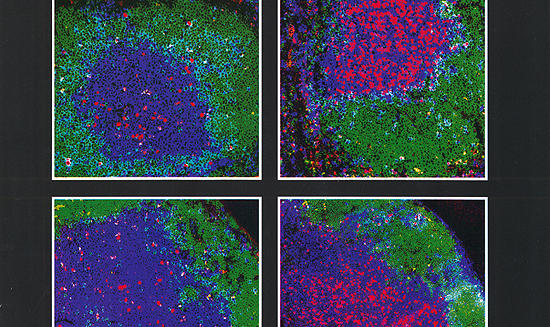
Memory T cell homing
Memory T cell homing Localization of distinct antigen-experienced CD8+ T cell populations in secondary lymphoid tissues. The figure shows four three-color confocal micrographs of cryostat sections of spleens (upper row) and peripheral lymph nodes (lower row) from recipient mice 24 hours after intravenous injection of TRITC labeled cytotoxic effector cells (left column) or central memory cells (right column). The sections were counterstained with anti-B220 (green) and anti-Thy-1.2 (blue) to localize B cell follicles and T cell areas, respectively. TRITC labeled homed donor cells are identified by red fluorescence.
False color photo showing platelet-mediated lymphocyte rolling via the interaction of P-selectin & Peripheral Node Addressin (PNAd)
Diacovo, T.G., Catalina, M.D., Siegelman, M.H., and von Andrian, U.H. Circulating activated platelets reconstitute lymphocyte homing and peripheral immunity in L-selectin deficient mice. J. Exp. Med. 187: 197-204, 1998. Diacovo, T.G., Puri, K.D., Springer, T.A., and von Andrian, U.H. Platelet-mediated lymphocyte delivery to high endothelial venules. Science 273: 252-255, 1996.
False color photo of a mouse peripheral lymph node displaying Peripheral Node Addressin (PNAd) staining with CarboxyFluorescein Succinimidyl Ester (CFSE) conjugated Meca-79 antibody
M'Rini, C., Cheng, G., Schweitzer, C., Cavanagh, L.L., Palframan, R.T., Mempel, T., Warnock, R.A., Lowe, J.B., Quackenbush, E.J., and von Andrian, U.H. A novel endothelial L-selectin ligand activity in lymph node medulla that is regulated by alpha (1,3)-fucosyltransferase-IV. J. Exp. Med. 198(9): 1301-1312, 2003. Stockton, B.M., Ardman, B., Cheng, G., Manjunath, N., and von Andrian, U.H. Negative regulation of T cell homing by CD43. Immunity 8: 373-381, 1998.
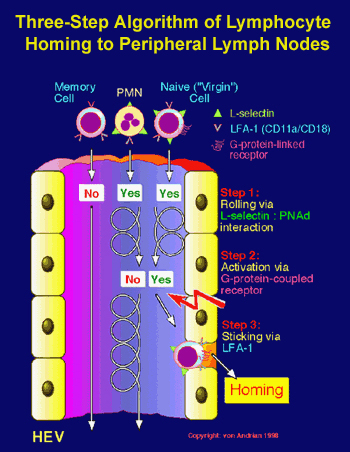
Three-step algorithm of lymphocyte homing to peripheral lymph nodes
Stein, J.V., Rot, A., Luo, Y., Narasimhaswamy, M., Nakano, H., Gunn, M.D., Matsuzawa, A., Quackenbush, E.J. Dorf, M.E., and von Andrian, U.H. The CC Chemokine Thymus-derived Chemotactic Agent 4 (TCA-4, Secondary Lymphoid Tissue Chemokine, 6Ckine, Exodus-2) Triggers Lymphocyte Function-associated Antigen 1-mediated Arrest of Rolling T Lymphocytes in Peripheral Lymph Node High Endothelial Venules. J. Exp. Med. 191: 61-75, 2000. Warnock, R.A., Askari, S., Butcher, E.C., and von Andrian, U.H. Molecular mechanisms of lymphocyte homing to peripheral lymph nodes. J. Exp. Med. 187: 205-216, 1998.